Cell Rich Zone Of Dental Pulp
Lymphocytes plasma cells and eosinophils are other cell types also common in dental pulp. Dental pulp stem cells DPSCs can be found within the cell rich zone of the dental pulp.
9 Dental Pulp Pocket Dentistry
The fibroblasts produce collagen fibers.
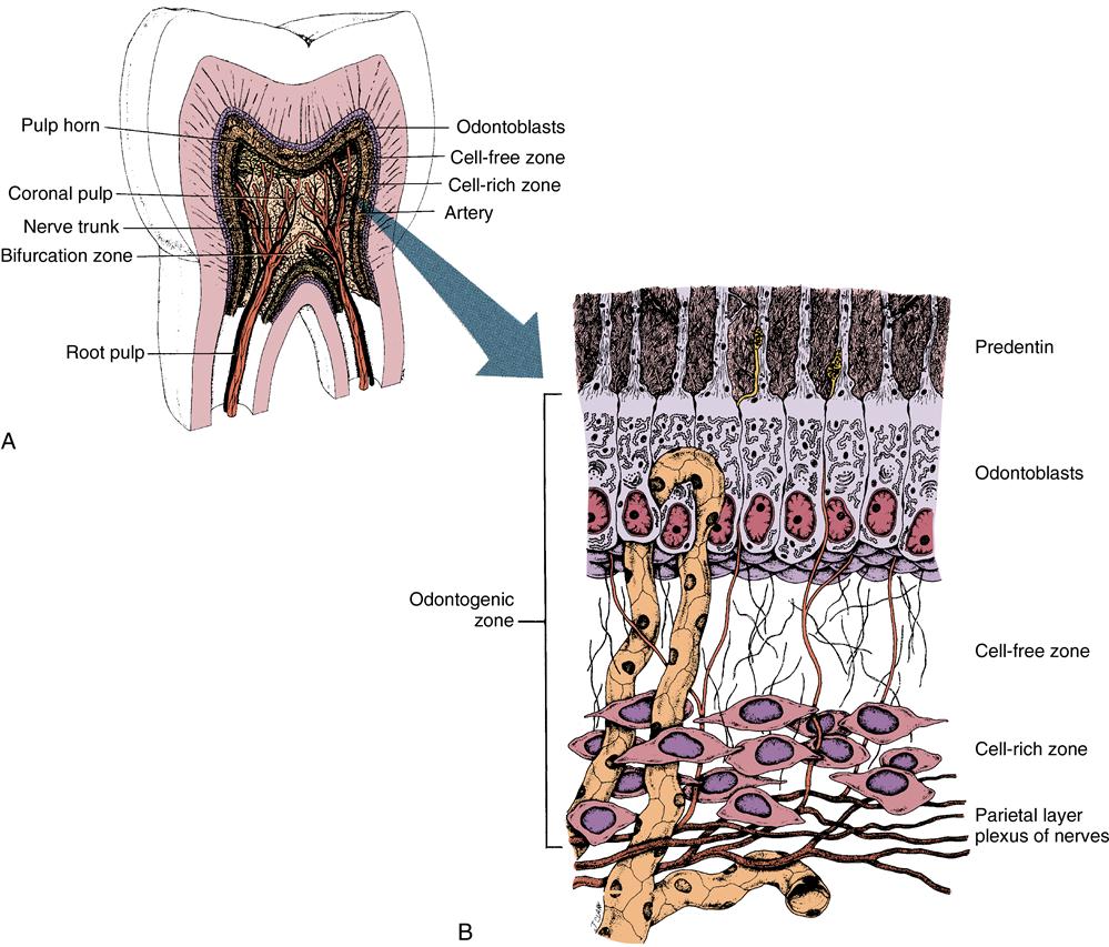
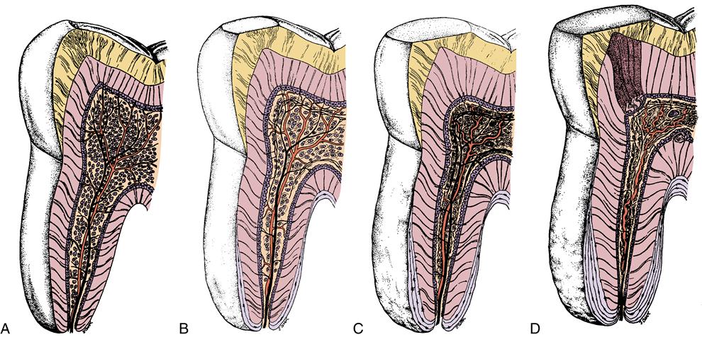
Cell rich zone of dental pulp. Zones Of Pulp The odontoblastic zone at the pulp periphery A Cell- Free Zone of Weil beneath the odontoblasts A Cell- Rich Zone Pulp Core 5. Cell-free zone of Weil appears as a space between the odontoblastic zone and the cell-rich zone. 5259646 PubMed - indexed for MEDLINE MeSH Terms.
The pulp cavity exhibits four zones as you progress from the dentin-pulp junction toward the center of the pulp cavity. Up to now it has been demonstrated that these cells are capable of producing bone tissue both in vitro and in vivo as well as a. Medial to the cell-rich zone is the deep pulp cavity 4 that contains subodontoblastic plexus of Raschkow.
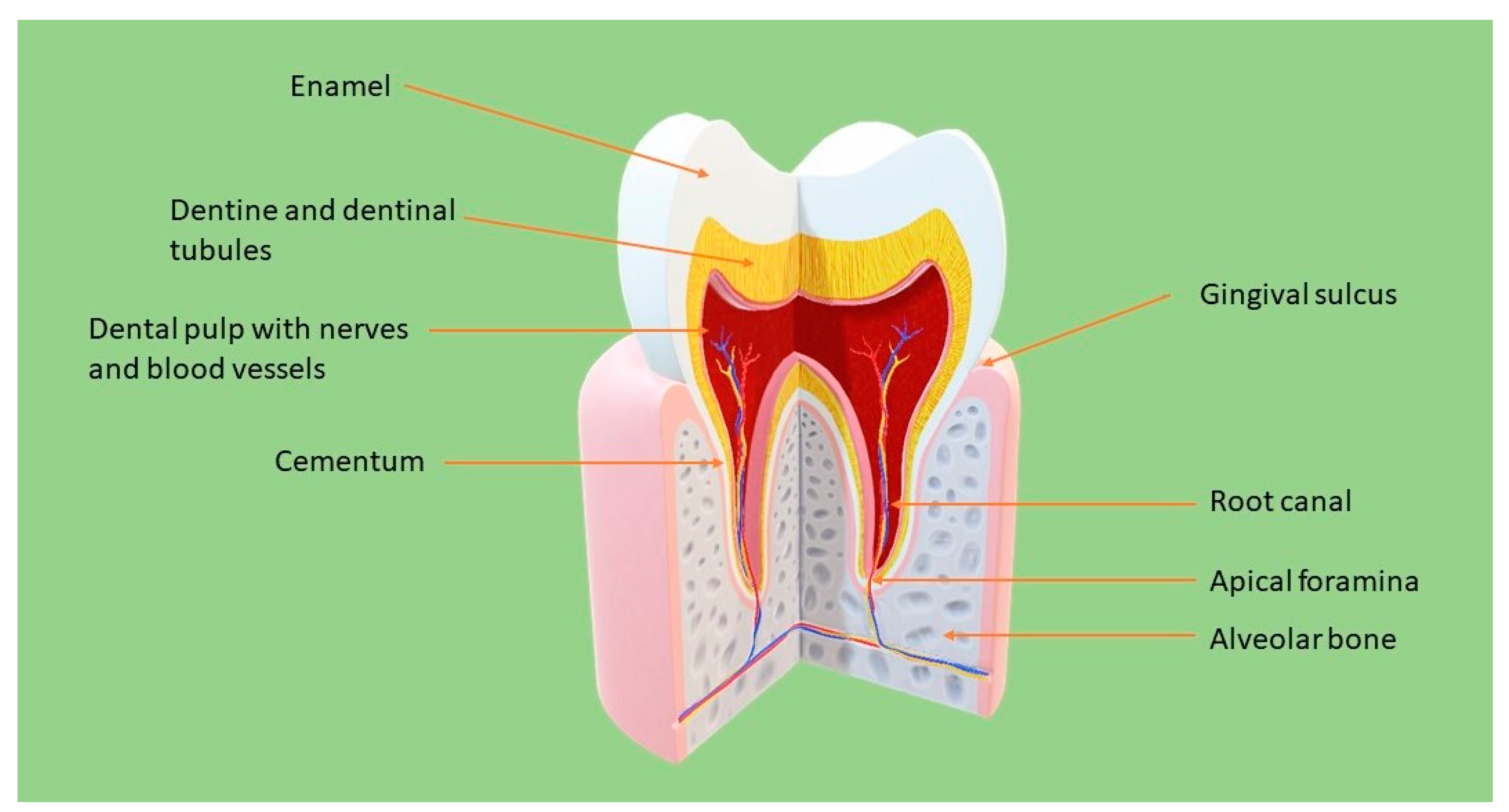
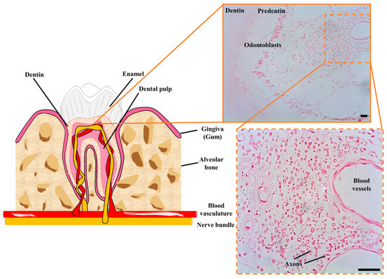
Parietal layer of nerves G. The dental pulp is the soft connective tissue of the tooth that supports the dentin. The surrounding apical zone an apical cell-rich zone is seen followed by the apical papilla mesenchyme.
Pulpal core which is in the center of the pulp chamber with many cells and an extensive vascular supply. Cellular organization in the subodontoblastic zone of the dental pulp. The intercellular substance.
The connective tissue of the pulp composed of. During early development there are also many young collagen fibers in this zone. 1 the odontoblast zone 2 cell-free zone basal layer of Weil 3 cell-rich zone and 4 the pulp core.
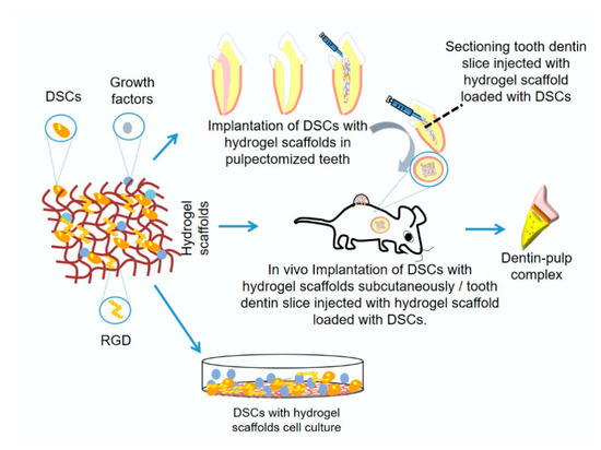
Dental pulp stem cells DPSCs can be found within the cell rich zone of dental pulp. Derived from the cells deeper in the pulp such as fibro-blasts in the cell-rich zone endothelial cells or pericytes of the blood vasculature which are differentiated upon the stimulation by tissue growth factor- 1011 The reparative dentine especially at the junctional zone between primary and. The cell rich zone is densely packed with undifferentiated mesenchymal cells and fibroblasts.
Lines the outer pulpal wall and consists of the cell bodies of odontoblast. Most abundant cell in pulp. Up to now it has been demonstrated that these cells are capable of producing bone tissue both in vitro and in vivo as well as a simil-dentin tissue in vitro.
These materials protect the pulp from noxious agents heat cold bacteria and stimulate the cell-rich zone of the pulp to lay down a bridge of reparative dentin. The cell-rich zone lies immediately under the cell-free zone and contains. Up to now two groups have studied these cells extensively albeit with different results.
Dentin formation usually starts within 30 days of the pulp capping there can be a delay in onset of dentin formation if the odontoblasts of the pulp are injured during cavity removal and is largely completed by 130 days. Typically long-lived cells lasting as long as the tooth remains vital. This layer is rich in cells which are fibroblasts and undifferentiated mesenchymal cells.
The cells of the pulp. Dental pulp stem cells DPSCs can be found within the cell rich zone of the dental pulp. Their embryonic origin from neural crests explains their multipotency.
The function of undifferentiated cells is that they become either fibroblasts or odontoblasts or macrophage according to need. Their embryonic origin from neural crests explains their multipotency. These cells cannot divide although it is currently thought that undifferentiated cells of the cell rich zone can be recruited to differentiate into odontoblasts to help repair exposed pulp.
The cells of the pulp include. Cell rich zone and core Activity corresponds to shape Producing and absorbing capabilities Undifferentiated ectomesenchymal cells Represent pool from which connective tissue cells of the pulp are derived Depending on the stimulus these cells may give rise either odontoblasts or fibroblasts. Their embryonic origin from neural crests explains their multipotency.
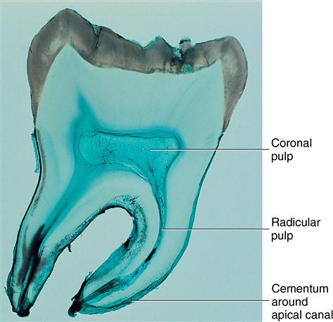
The radicular dental pulp is located in the root and this is the place where pulp cells differentiate into odontoblasts and in the sub-odontoblastic layer the so-called Hoehls layer Figures 91013-14. A cell-free zone is not present in developing teeth but becomes prominent in the coronal pulp after development. Tall columnar odontoblasts are arranged in Palisading pattern forming a single layer in the Peripheral area of the pulp.
CELL FREE ZONE OF WEIL. Which contains fibroblasts and. Cell rich zone.
Period and mode of development of the cell-rich layer in rat molar pulps. Dentin Predentin Odontoblasts Layer Cell rich Zone Cell free Zone Pulp Proper 6. This area is lined peripherally by a specialized odontogenic area which has four layers from innermost to outermost.

Immunohistochemical Staining Of Mesenchymal Markers In Human Dental Download Scientific Diagram
Plos One Teneurin 2 Presence In Rat And Human Odontoblasts
Polymers Free Full Text Hydrogels And Dentin Pulp Complex Regeneration From The Benchtop To Clinical Translation Html
Molecules Free Full Text Ils And Mmps Levels In Inflamed Human Dental Pulp A Systematic Review Html

Characterization Of Human Dental Pulp Stem Cell Distribution A Download Scientific Diagram

Pin On Oral Histology

Schematic Representation Of The Suggested Relationship Between Notch Download Scientific Diagram

Immunohistochemical Staining Of Neural Markers In Human Dental Pulp Download Scientific Diagram

Control And Irradiated Samples Exhibiting Preservation Of The Dental Download Scientific Diagram

Photomicrographs Of Histologic Sections Of Dental Pulp All Stained Download Scientific Diagram
Dental Pulp Dentin Capillary


Dental Pulp Autotransplantation A New Modality Of Endodontic Regenerative Therapy Follow Up Of 3 Clinical Cases Journal Of Endodontics
9 Dental Pulp Pocket Dentistry

Tooth Structure And Dental Tissues With The Respective Stem Cell Download Scientific Diagram

Uncovered Dental Surgery Assistant Toothpastedispenser Dentalimplantscostpeople Dental Hygiene School Dental Hygienist Dental Hygiene Student
9 Dental Pulp Pocket Dentistry
Oral Histology Digital Lab Dental Pulp Zones Of The Pulp Image 4

Ijms Free Full Text Advances And Perspectives In Dental Pulp Stem Cell Based Neuroregeneration Therapies Html

Schematic Drawing Of The Peripherical Part Of The Dental Pulp D Download Scientific Diagram
Post a Comment for "Cell Rich Zone Of Dental Pulp"